
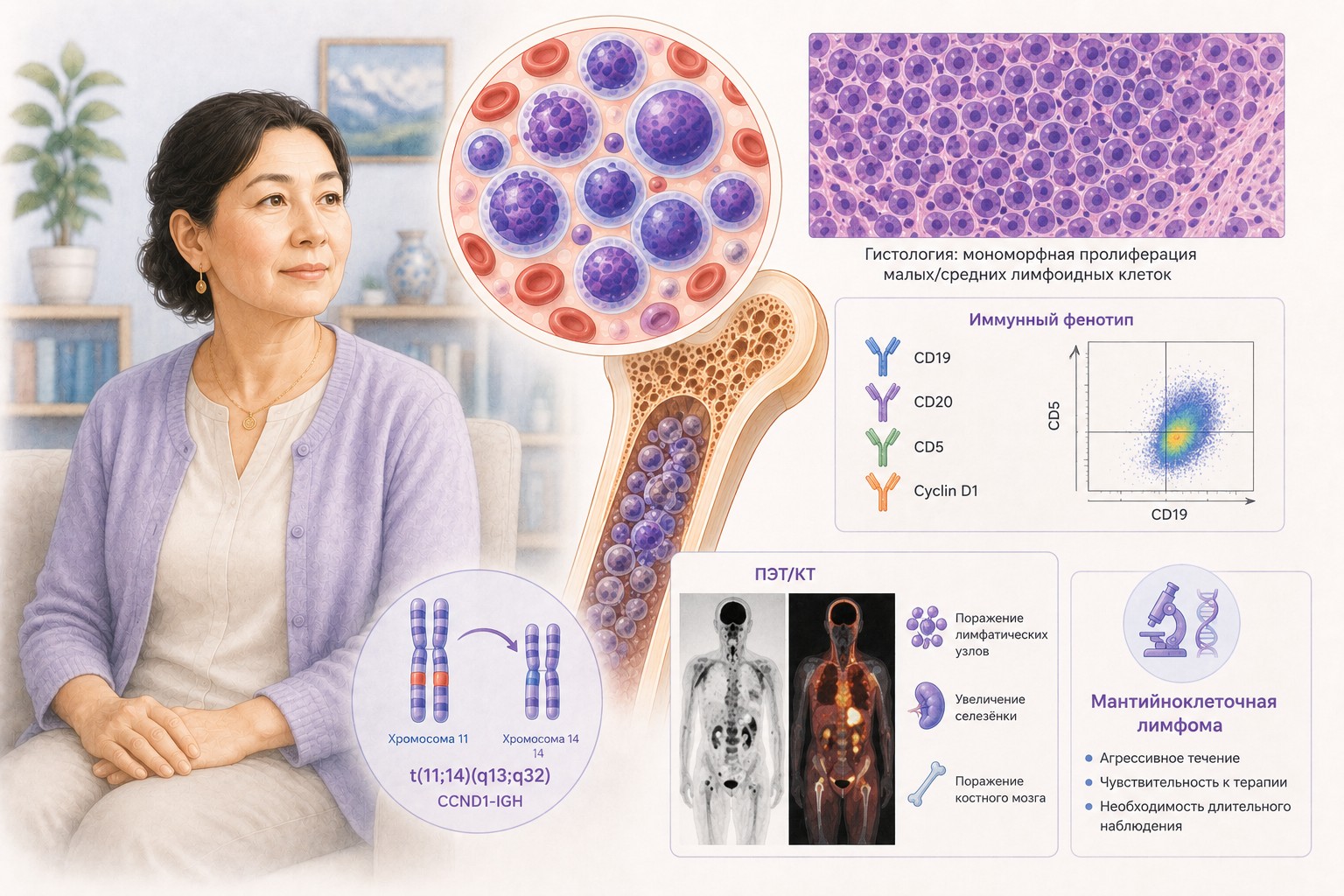
Mantle Cell Lymphoma (MCL): Key Information for Patients
Based on NCCN Guidelines for Patients® — Mantle Cell Lymphoma, 2025 (NCCN Clinical Practice Guidelines in Oncology, B-Cell Lymphomas Version 2.2025 — February 10, 2025)
What Is Mantle Cell Lymphoma
Mantle cell lymphoma (MCL) is a rare but treatable cancer that develops from B lymphocytes located in the mantle zone — the outer ring of cells surrounding the germinal center inside lymph nodes. MCL occurs when these cells develop a genetic change (translocation t(11;14)) that causes overproduction of a protein called cyclin D1, which drives uncontrolled cell growth.
MCL is usually an aggressive, fast-growing cancer, but in some patients it follows an indolent (slow-growing) course. The growth pattern significantly influences treatment choices.
Most people are diagnosed with advanced-stage MCL (stages 3–4), as the disease typically spreads to multiple areas before causing noticeable symptoms. MCL commonly involves not only lymph nodes but also the spleen, liver, bone marrow, bloodstream, and gastrointestinal (GI) tract — more so than other lymphomas.
MCL is usually a lifelong disease with no cure in most cases, but modern treatments can achieve remission and significantly extend life.
Symptoms
- Painless, swollen lymph nodes (neck, armpit, groin) — most common
- B symptoms: fever, drenching night sweats, unexplained weight loss
- Fatigue and weakness
- Enlarged spleen or liver (fullness, abdominal discomfort)
- GI tract symptoms: diarrhea, bloody stools, abdominal pain
- Frequent infections
Diagnostic Workup
Required tests:
- Full medical history including B symptoms
- Physical exam: lymph nodes, spleen, liver size; performance status
- CBC with differential; comprehensive metabolic panel; LDH; uric acid; beta-2 microglobulin
- Hepatitis B and C testing (before treatment)
- CT or PET/CT scan of chest, abdomen, and pelvis (with contrast; neck if needed)
- Echocardiogram or MUGA scan — if anthracycline-based chemotherapy is planned
Biopsy (essential for diagnosis):
- Excisional or incisional lymph node biopsy preferred; core needle biopsy if lymph node is inaccessible
- Bone marrow biopsy ± aspirate — when indicated
- GI endoscopy/colonoscopy — when GI involvement is suspected (more common in MCL than other lymphomas)
- Lumbar puncture — if CNS involvement is suspected
Pathology panel:
| Marker | MCL finding |
|---|---|
| Cyclin D1 | Positive (hallmark) |
| CD5, CD20 | Positive |
| SOX11 | Usually positive (absent in indolent MCL) |
| CD200, LEF1 | Negative (absent in MCL) |
| t(11;14) translocation | Present (FISH or karyotype) |
| Ki-67 | High = fast-growing; quantifies proliferation |
Molecular biomarker testing:
- TP53 mutation — critical: TP53-mutated MCL is high-risk, grows rapidly, responds poorly to standard treatment. NCCN strongly recommends clinical trial for TP53-mutated MCL.
- Additional chromosomal abnormalities assessed for prognosis
Blastoid variant: rare subtype with large, rapidly dividing cells; grows faster and is harder to treat; may appear at diagnosis or develop later.
Staging
MCL is staged using the Ann Arbor system:
- Stage 1: disease in one lymph node region
- Stage 2: disease in ≥2 lymph node regions on the same side of the diaphragm
- Stage 3–4: advanced — multiple lymph node regions, bone marrow, other organs
MCL is rarely diagnosed at stage 1–2 (early disease). Almost all patients have advanced-stage disease at diagnosis.
Treatment
Slow-Growing (Indolent) MCL
Asymptomatic patients with slow-growing MCL (often SOX11-negative; disease confined to bone marrow/blood) may be managed with active surveillance (watch and wait) — regular monitoring without treatment. Check-ups every 2–3 months while stable. If symptoms develop or growth accelerates, treatment begins (re-evaluate for TP53 mutation).
Early-Stage MCL (Stages 1–2)
Rarely diagnosed. Options:
- Radiation therapy (ISRT) alone
- Chemoimmunotherapy ± radiation
- Active surveillance in select cases
Follow-up after complete response: physical exam + labs every 3–6 months for 5 years, then annually; CT scan ≤2×/year for 2 years.
Advanced MCL (Stages 3–4), Fast-Growing
First-line therapy proceeds in up to three phases:
Phase 1: Primary treatment — goal is maximum tumor reduction
For patients who can tolerate aggressive therapy:
| Regimen | Components |
|---|---|
| LYMA (preferred) | RDHAP → RCHOP |
| Nordic (preferred) | Dose-intensified RCHOP/RDHAP alternating with high-dose cytarabine |
| BR → R + HD-Ara-C (preferred) | Bendamustine + rituximab → rituximab + high-dose cytarabine |
| TRIANGLE (preferred) | RCHOP + ibrutinib alternating with RDHAP |
| HyperCVAD+R (other) | Cyclophosphamide, vincristine, doxorubicin, dexamethasone + rituximab |
| RBAC (other) | Rituximab, bendamustine, cytarabine |
For patients who cannot tolerate aggressive therapy:
| Regimen | |
|---|---|
| ABR (preferred) | Acalabrutinib + bendamustine + rituximab |
| BR (preferred) | Bendamustine + rituximab |
| VR-CAP (preferred) | Bortezomib + rituximab + cyclophosphamide + doxorubicin + prednisone |
| RCHOP (preferred) | |
| LR (preferred) | Lenalidomide + rituximab |
| AR (other) | Acalabrutinib + rituximab |
Phase 2: Additional (consolidation) treatment — if complete response to primary therapy:
- Targeted therapy with a BTK inhibitor (acalabrutinib, ibrutinib, or zanubrutinib) — now preferred over autologous stem cell transplant (HDT/ASCR) for most patients
- If only partial response: BTK inhibitor (if not previously used) or chemoimmunotherapy
- Clinical trial
Phase 3: Maintenance treatment — to prevent or delay relapse:
- Rituximab IV every 8 weeks for 2–3 years
- If aggressive primary therapy was used: rituximab + BTK inhibitor for 2 years
TP53-Mutated MCL
NCCN strongly recommends clinical trial as first choice.
If no trial available:
- Zanubrutinib + obinutuzumab + venetoclax (new combination)
- Aggressive therapy capable patients: TRIANGLE regimen → maintenance rituximab q8w × 3 years + ibrutinib × 2 years
- Less aggressive: options from less-aggressive list → rituximab maintenance
Relapsed and Refractory MCL
Second-line (preferred):
- Acalabrutinib (continuous)
- Zanubrutinib (continuous)
- Lenalidomide + rituximab
Second-line (other recommended): ibrutinib ± rituximab
Second-line (certain cases): bendamustine + rituximab; DHA-platinum-rituximab; GemOx-R; ibrutinib + venetoclax; RBAC; venetoclax ± rituximab
Third-line and beyond — Guide 6 criteria:
- BTK inhibitor: only if not previously used, OR switching to pirtobrutinib (non-covalent BTK inhibitor, active after covalent BTK inhibitor failure)
- CAR T-cell therapy (brexucabtagene autoleucel/Tecartus or lisocabtagene maraleucel/Breyanzi): only after chemoimmunotherapy AND prior BTK inhibitor
- Bispecific antibody (glofitamab-gxbm/Columvi): only after CAR T + pirtobrutinib, or if CAR T-cell therapy is not an option
- Autologous HCT (HDT/ASCR): only in select cases, only if no prior transplant
- Allogeneic HCT: only if MCL is in remission AND CAR T-cell therapy has already been done
Clinical trial is strongly encouraged at all stages of relapsed/refractory disease.
Follow-up: every 3–6 months with physical exam, blood tests, and CT imaging.
Supportive Care
Common side effects of MCL treatments: fatigue, nausea, vomiting, diarrhea, headache, low blood counts, constipation, infection, fever/chills, decreased appetite, rash, oral mucositis, shortness of breath, arrhythmia, hair loss, peripheral neuropathy (tingling/numbness in hands and feet).
Low-dose palliative radiation can relieve bone pain and other localized symptoms without treating the underlying lymphoma systemically.
Advance care planning is appropriate for all patients — not only those at end of life. Early conversations about future preferences reduce stress and improve quality of life.
Key Messages for Patients
- MCL is usually diagnosed at an advanced stage, but effective treatments exist. Modern therapies have significantly improved outcomes compared to even a decade ago.
- Know your TP53 status — TP53 mutation changes treatment strategy fundamentally. If positive, a clinical trial is the first priority; if unavailable, a specific triple combination is recommended.
- Slow-growing MCL without symptoms does not require immediate treatment — active surveillance is a valid evidence-based approach.
- The three-phase first-line treatment (primary → consolidation → maintenance) is complex. Discuss each phase with your care team and understand the role of BTK inhibitors in consolidation and rituximab in maintenance.
- HDT/ASCR (autologous transplant) is no longer the default consolidation for most MCL patients — BTK inhibitors have taken this role in current guidelines.
- When MCL relapses, pirtobrutinib offers a new option specifically for patients who have already received other BTK inhibitors.
- CAR T-cell therapy (Tecartus, Breyanzi) is a potent option for multiply relapsed MCL but requires prior chemoimmunotherapy AND prior BTK inhibitor.
- Clinical trials should be considered at every stage — MCL is a focus of active research with new treatments emerging regularly.
- Because MCL is rare and complex, seek care at or consultation with a center specializing in lymphomas.
Source: NCCN Guidelines for Patients® — Mantle Cell Lymphoma, 2025 (based on NCCN Clinical Practice Guidelines in Oncology, B-Cell Lymphomas Version 2.2025 — February 10, 2025). National Comprehensive Cancer Network® (NCCN®).
Full original document available free of charge: NCCN.org/patientguidelines
This material is for informational purposes only and does not replace consultation with a treating physician.
Download PDF
pa_d6b0548a-5986-4f0e-9350-2b2f47188171_1778339976062.pdf