
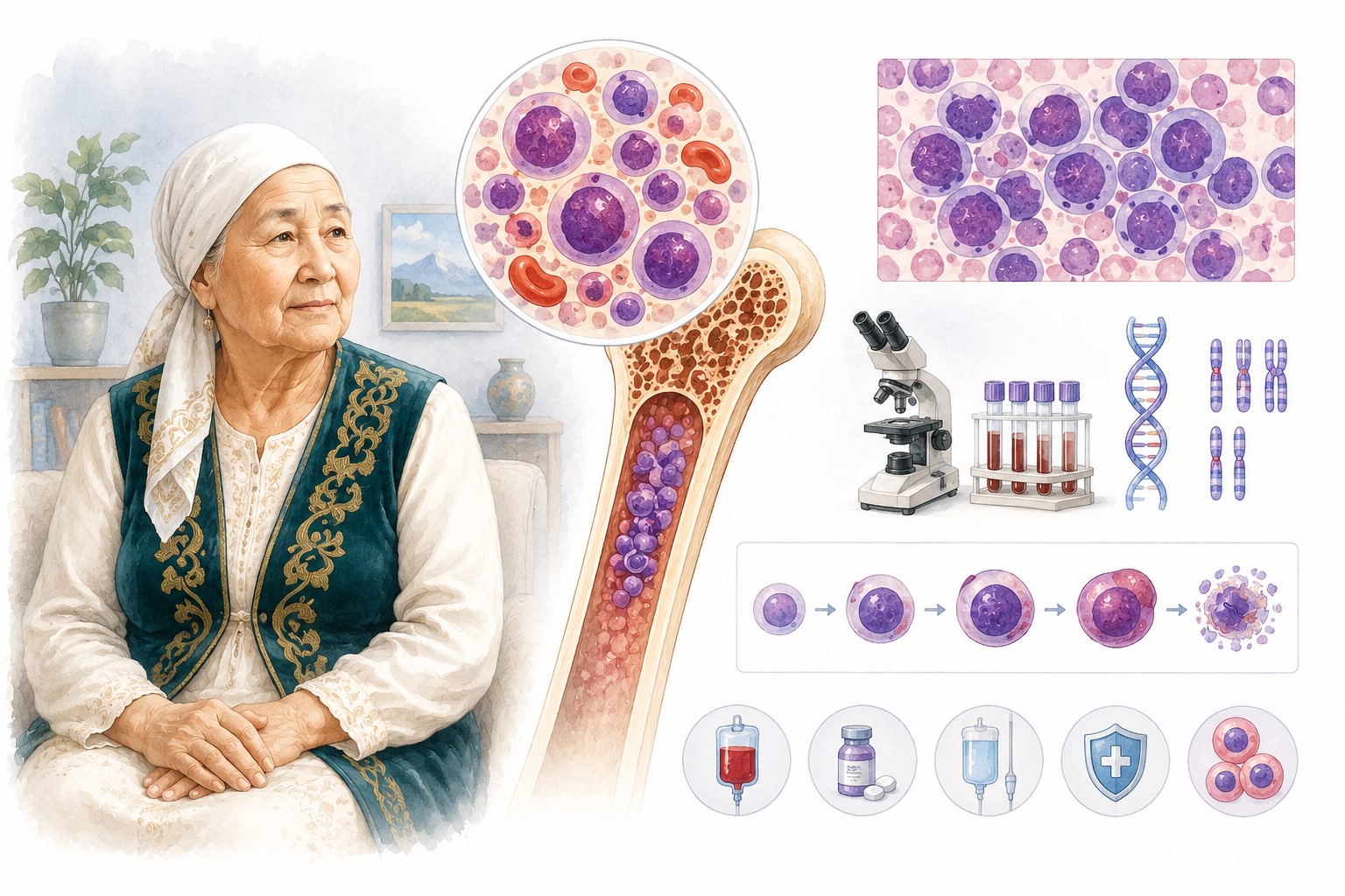
Acute Myeloid Leukemia (AML): Key Information for Patients
Based on NCCN Guidelines for Patients® — Acute Myeloid Leukemia, 2026 (NCCN Clinical Practice Guidelines in Oncology, Version 3.2026 — November 24, 2025)
What Is AML
Acute myeloid leukemia (AML) is a fast-growing blood cancer that starts in the bone marrow. Abnormal changes prevent very immature white blood cells called myeloblasts (blasts) from developing into mature blood cells. Blasts accumulate in the bone marrow and blood, crowding out healthy red blood cells, white blood cells, and platelets. This causes serious health problems and is fatal if untreated.
AML has no traditional stages. Instead, it is classified by specific gene mutations and chromosomal changes within the leukemia cells, which guide treatment decisions.
This guide also covers two related diseases included in the same NCCN document:
- Acute promyelocytic leukemia (APL) — a rare, highly curable AML subtype defined by the PML::RARA fusion gene
- Blastic plasmacytoid dendritic cell neoplasm (BPDCN) — a very rare, aggressive blood cancer affecting skin, lymph nodes, and bone marrow
Part 1: AML
Symptoms
- Extreme fatigue, weakness, pale skin
- Frequent or severe infections, fever
- Easy bruising, bleeding gums, nosebleeds, slow-healing cuts
- Shortness of breath
- Bone or joint pain
- Unexplained weight loss or loss of appetite
- Swollen lymph nodes or enlarged spleen
Diagnostic Workup
Required tests:
- Medical and family history; physical exam; performance status assessment; distress screening
- CBC with differential; comprehensive metabolic panel; LDH; uric acid; B12, folate; iron
- Blood clotting tests (coagulation panel)
- Bone marrow aspirate and biopsy — essential for diagnosis; confirms ≥20% blasts in bone marrow or blood
- AML biomarker and genetic testing (on bone marrow or blood sample): karyotype, FISH, NGS, RT-PCR — to identify gene mutations and chromosomal abnormalities
- HLA typing — if transplant may be needed
- ECG and echocardiogram — to assess heart before anthracycline-based treatment
Imaging when indicated: FDG-PET/CT (if extramedullary disease suspected); brain CT without contrast (if CNS bleeding suspected); brain MRI with contrast (if leukemic meningitis suspected)
Lumbar puncture: if CNS involvement is suspected
Fertility counseling: before any systemic therapy that could affect fertility
Key Genetic Mutations and Their Significance
| Mutation / Change | Significance |
|---|---|
| FLT3-ITD or FLT3-TKD | Targeted by midostaurin, quizartinib, gilteritinib, sorafenib |
| IDH1 mutation | Targeted by ivosidenib, olutasidenib |
| IDH2 mutation | Targeted by enasidenib |
| KMT2A rearrangement | Targeted by revumenib |
| NPM1 mutation | Targeted by revumenib, ziftomenib |
| CD33-positive | Targeted by gemtuzumab ozogamicin |
| Core binding factor (CBF): t(8;21) or inv(16) | Favorable risk; responds well to cytarabine-based regimens |
| TP53 mutation | Poor risk; clinical trial strongly recommended |
Risk Groups (ELN 2022/2024)
| Risk group | Example features |
|---|---|
| Favorable | t(8;21)/RUNX1::RUNX1T1; inv(16)/CBFB::MYH11; mutated NPM1 without FLT3-ITD; CEBPA bZIP mutation |
| Intermediate | Mutated NPM1 with FLT3-ITD; wild-type NPM1 with FLT3-ITD; t(9;11)/MLLT3::KMT2A |
| Poor | TP53 mutation; KMT2A rearrangement; complex/monosomal karyotype; ASXL1, BCOR, EZH2, RUNX1, SRSF2, STAG2, TP53 and other adverse mutations; -5 or del(5q); -7; -17/abn(17p) |
Treatment Phases
AML is always treated in phases:
- Induction (remission induction): destroy blasts and achieve complete remission (CR); takes ~4–6 weeks for bone marrow recovery
- Consolidation (post-remission therapy): eliminate any surviving blasts; deepen and prolong remission
- Maintenance (for some patients): prolong remission; continues for months to years
- Surveillance: monitoring after remission for signs of relapse (CBC every 1–3 months for 2 years, then every 3–6 months for up to 5 years)
Measurable residual disease (MRD): very sensitive tests (PCR) may detect leukemia cells even in apparent remission; MRD positivity guides decisions about transplant or additional therapy.
Intensive Induction
For patients who can tolerate intensive chemotherapy:
Favorable- and intermediate-risk AML:
- Preferred: Standard 7+3 (7 days cytarabine + 3 days daunorubicin or idarubicin); for CBF-AML: 7+3 + gemtuzumab ozogamicin if CD33+
- Other recommended: FLAG-IDA ± gemtuzumab ozogamicin; CLAG-M
- FLT3-ITD: 7+3 + midostaurin or quizartinib
- FLT3-TKD: 7+3 + midostaurin
Poor-risk AML (therapy-related, antecedent MDS/CMML, or MDS-related chromosomal changes):
- Preferred: CPX-351/Vyxeos (age ≥60); standard 7+3 (age ≤60)
- Other: decitabine + venetoclax; azacitidine + venetoclax; CLIA; FLAG-IDA ± venetoclax
Poor-risk AML (other):
- Clinical trial is recommended
- Other: 7+3; CPX-351; FLAG-IDA; decitabine or azacitidine + venetoclax; CLAG-M
TP53-mutated or del(17p) AML: clinical trial
Less Intensive Induction
For patients who cannot tolerate intensive chemotherapy (due to age, health, or personal preference). Less intensive therapy can still achieve complete remission. It is typically continued indefinitely as long as it controls the disease.
AML with IDH1 mutation:
- Preferred: azacitidine or decitabine + venetoclax; azacitidine + ivosidenib
- Other: ivosidenib; LDAC + venetoclax; azacitidine or decitabine alone; olutasidenib
AML without IDH1 mutation:
- Preferred: azacitidine or decitabine + venetoclax
- Other: cladribine + LDAC + venetoclax; LDAC + venetoclax; azacitidine or decitabine alone; LDAC + glasdegib; LDAC alone
- FLT3 mutation: gilteritinib ± azacitidine
- IDH2 mutation: enasidenib ± azacitidine
- CD33+: gemtuzumab ozogamicin
After Induction
- Complete response: cytarabine-based consolidation, continuation of prior therapy, or allogeneic HCT; clinical trial if available
- Less than complete response / progression: chemotherapy, targeted therapy, clinical trial, HCT, or best supportive care
Maintenance:
- After intensive induction: azacitidine may be used
- After less intensive induction: continue same therapy
- FLT3 mutation history: targeted therapy maintenance
- After HCT: individualized
Relapsed and Refractory AML
Relapsed AML: disease returns after remission. Refractory AML: disease does not respond to treatment or worsens during treatment.
Biomarker testing (NPM1, IDH1, IDH2, FLT3) should be repeated at each relapse — mutations may change.
Treatment options:
- Clinical trial (strongly preferred)
- Targeted therapy based on mutation (see table above) or chemotherapy → then HCT
- Best supportive care
Targeted therapy by mutation at relapse:
| Mutation | Preferred agents |
|---|---|
| FLT3-ITD | Gilteritinib; HMA + sorafenib; quizartinib |
| FLT3-TKD | Gilteritinib |
| IDH1 | Ivosidenib; olutasidenib |
| IDH2 | Enasidenib |
| KMT2A rearrangement | Revumenib |
| NPM1 | Revumenib; ziftomenib |
| CD33+ | Gemtuzumab ozogamicin |
Chemotherapy options for relapsed/refractory AML (intensive): CLAG ± mitoxantrone/idarubicin; cytarabine ± anthracycline; FLAG-IDA ± venetoclax; etoposide + cytarabine ± mitoxantrone; clofarabine ± cytarabine/idarubicin; CLIA + venetoclax
Less intensive: azacitidine or decitabine ± venetoclax; LDAC + venetoclax
Allogeneic HCT
The only potentially curative treatment for many AML subtypes. Replaces diseased bone marrow with donor stem cells. Requires HLA typing to find a matched donor.
Process: conditioning chemotherapy (± radiation) to destroy leukemia → donor stem cell infusion → engraftment (2–4 weeks, during which patient is vulnerable to infections) → new healthy bone marrow forms.
Goal: the new immune system recognizes residual leukemia as foreign (graft-versus-leukemia effect) and destroys it.
Key side effects: febrile neutropenia, infections, bleeding, graft-versus-host disease (GVHD) — donor cells attack normal tissues. GVHD is treatable but serious.
Supportive Care
Febrile neutropenia: fever with low neutrophils is a medical emergency; requires immediate IV antibiotics.
High blast count (hyperleukocytosis): apheresis or hydroxyurea used to rapidly reduce white blood cell count before treatment.
Tumor lysis syndrome (TLS): dangerous electrolyte disturbances from rapid cell death during treatment; prevented by hydration, allopurinol/rasburicase.
Differentiation syndrome: life-threatening cytokine release from leukemia cells responding to treatment; symptoms include fever, limb swelling, difficulty breathing, weight gain, skin rash; treated with steroids and hydroxyurea.
Blood transfusions: red blood cell transfusions for anemia; platelet transfusions for thrombocytopenia. Growth factors (G-CSF) to boost white blood cells.
Cytarabine-specific toxicities: cerebellar ataxia (movement disorders); eye inflammation (prevented with saline/steroid eye drops).
Part 2: Acute Promyelocytic Leukemia (APL)
APL is a rare subtype (~10% of AML). APL is curable more often than any other AML subtype — but it is a medical emergency: it causes life-threatening bleeding that can be fatal within hours of diagnosis if not treated immediately.
Defining feature: PML::RARA fusion gene from translocation t(15;17); confirmed by FISH or RT-PCR.
Start ATRA immediately if APL is suspected — even before confirmation. ATRA stops the coagulopathy. If tests rule out APL, ATRA is stopped.
Risk groups:
- Low risk: WBC ≤10 × 10⁹/L at diagnosis
- High risk: WBC >10 × 10⁹/L at diagnosis
Treatment:
Low risk:
- Preferred induction: ATRA + arsenic trioxide (ATO)
- Consolidation: ATRA + ATO
- If ATO not available: ATRA + idarubicin or gemtuzumab ozogamicin
High risk (no cardiac issues):
- Preferred induction: ATRA + ATO + idarubicin or gemtuzumab ozogamicin
- Other: ATRA + daunorubicin + cytarabine; ATRA + idarubicin
High risk with cardiac issues (low ejection fraction or prolonged QTc):
- ATRA-based regimen adjusted to cardiac status
Maintenance: possible; individualized.
Monitoring: PCR testing of bone marrow/blood after consolidation; no treatment during monitoring. Watch for return of PML::RARA gene.
Relapse: early (<6 months) vs. late (≥6 months) determines re-treatment approach. Goal is remission. After remission: HCT, ATO, or clinical trial.
APL-specific supportive care:
- Coagulopathy: correct with platelet transfusions, cryoprecipitate, fresh frozen plasma — ongoing until coagulopathy resolves
- Differentiation syndrome: fever, breathing difficulties, fluid accumulation; steroids are treatment; hydroxyurea if rising WBC
- Arsenic trioxide cardiac monitoring: ECG required throughout treatment; arsenic prolongs QTc interval; electrolytes monitored
Part 3: Blastic Plasmacytoid Dendritic Cell Neoplasm (BPDCN)
BPDCN is a very rare, aggressive blood cancer of immature plasmacytoid dendritic cells. It is often misdiagnosed because of its varied presentation. Treatment requires a multidisciplinary team including a dermatologist.
Typical presentation: dark purple skin lesions (may look like bruises or a rash); enlarged lymph nodes; fatigue; abdominal pain from spleen involvement. Can involve CNS, blood, and bone marrow.
Diagnosis: skin biopsy (most common), lymph node biopsy, or bone marrow biopsy; hematopathologist review essential; biomarker/genetic testing (TET2, ASXL1, TP53, IDH2, and others).
Treatment (intensive):
- Preferred: tagraxofusp-erzs (Elzonris) — targets CD123 protein on BPDCN cells; first cycle must be given inpatient with ≥24-hour monitoring; major risk: capillary leak syndrome (fluid leaks from blood vessels → dangerous drop in blood pressure, organ failure, death)
- Chemotherapy alternatives: HyperCVAD; cytarabine + idarubicin/daunorubicin; CHOP
- After complete response: continue tagraxofusp-erzs until progression, or proceed to HCT
Treatment (non-intensive):
- Localized skin disease: radiation or surgery
- Systemic disease: venetoclax + azacitidine or decitabine; systemic steroids; supportive care
Relapsed/refractory BPDCN:
- Clinical trial (preferred)
- Tagraxofusp-erzs if not previously used
- Chemotherapy or radiation if not previously used
- HMA + venetoclax
CNS prophylaxis: intrathecal chemotherapy for all patients with CNS disease or suspected CNS disease at diagnosis.
Surveillance: CBC every 1–3 months for 2 years, then every 3–6 months for up to 5 years; bone marrow biopsy as needed; FDG-PET/CT if extramedullary disease was present.
Key Messages for Patients
- AML is serious and progresses quickly — treatment at a specialized center with AML expertise is essential. Seek care immediately.
- Genetic and biomarker testing drives every treatment decision. Know your mutation profile — FLT3, IDH1, IDH2, NPM1, TP53, KMT2A are all targetable or prognostically critical.
- Clinical trials are strongly recommended at every stage, especially for poor-risk AML, relapsed/refractory disease, and BPDCN.
- Targeted therapies have transformed AML treatment — many patients who previously had no options now have effective targeted agents. Ask whether your mutation qualifies you.
- APL is a medical emergency: if APL is suspected, ATRA must be started immediately to prevent fatal bleeding, even before genetic confirmation.
- Differentiation syndrome can occur with APL treatment and some AML targeted therapies — report any fever, swelling, or breathing difficulty immediately.
- Fertility preservation options should be discussed before starting any therapy.
- MRD status after induction guides whether transplant is needed — ask your care team about MRD testing and what your result means.
- Late effects — heart damage, cognitive effects, secondary cancers — can occur years after treatment. Follow-up care is lifelong.
Source: NCCN Guidelines for Patients® — Acute Myeloid Leukemia, 2026 (based on NCCN Clinical Practice Guidelines in Oncology, Version 3.2026 — November 24, 2025). National Comprehensive Cancer Network® (NCCN®).
Full original document available free of charge: NCCN.org/patientguidelines
This material is for informational purposes only and does not replace consultation with a treating physician.
Download PDF
pa_c112fac5-9a55-4382-a2c1-03278a3ad9b2_1778390699523.pdf