
Waldenström Macroglobulinemia (WM): Key Information for Patients
Based on NCCN Guidelines for Patients® — Waldenström Macroglobulinemia/Lymphoplasmacytic Lymphoma, 2025 (NCCN Clinical Practice Guidelines in Oncology, Version 3.2025 — February 6, 2025)
What Is Waldenström Macroglobulinemia
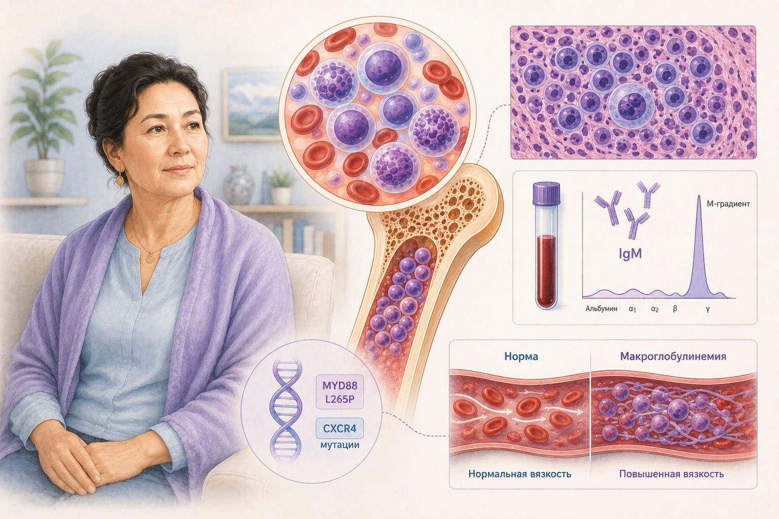
Waldenström macroglobulinemia (WM) is a rare, slow-growing blood cancer that begins in B lymphocytes — a type of white blood cell. In WM, a single immune cell undergoes a genetic change and gives rise to abnormal lymphocytes, plasma cells, and lymphoplasmacytic cells (cells intermediate between the two). Together, these are called WM cells.
WM cells do two harmful things:
- They crowd out healthy blood cells in the bone marrow, causing anemia, infections, and bleeding.
- They produce large amounts of a single abnormal antibody — immunoglobulin M (IgM) — the largest of the five types of immunoglobulins. When excess IgM builds up in the blood, it can thicken the blood, a dangerous condition called hyperviscosity syndrome.
WM and lymphoplasmacytic lymphoma (LPL) are closely related. LPL is the cancer; WM is the most common type of LPL (defined by the presence of high IgM levels). Treatment of both is the same.
There is currently no cure for WM, but many effective therapies can control the disease, relieve symptoms, and provide years of good quality of life.
Symptoms
Many people with WM have no symptoms for years and never need treatment. When symptoms occur, they may include:
- Fatigue and weakness (most common)
- Fever, night sweats, unexplained weight loss
- Numbness, tingling, or pain in hands and feet (peripheral neuropathy)
- Swollen lymph nodes, enlarged spleen or liver (swollen abdomen)
- Easy bruising, nosebleeds, or bleeding gums
- Headaches, dizziness, blurred or double vision, confusion
- Shortness of breath
- Sensitivity to cold
WM complications and their specific symptoms:
| Complication | What it is | Key symptoms |
|---|---|---|
| Hyperviscosity syndrome | Blood thickening from excess IgM | Nosebleeds, headache, blurred vision, dizziness, chest pain |
| Anemia | Low red blood cells | Fatigue, dizziness, rapid heartbeat, pallor |
| Peripheral neuropathy | IgM-mediated nerve damage | Numbness, tingling, burning, weakness in hands/feet |
| Cold agglutinin disease | RBC destruction in cold | Fatigue, blue/painful fingers or toes, jaundice |
| Cryoglobulinemia | Vessel blockage in cold | Purple skin lesions, joint pain, hand/foot color changes |
| Amyloidosis | Abnormal protein buildup in organs | Weight loss, leg/ankle swelling, numbness, breathlessness |
Diagnostic Workup
Required tests:
- Full history and physical examination (lymph node palpation, spleen/liver size)
- CBC (typically shows anemia; low white blood cells and platelets possible)
- Serum immunoglobulin levels — characteristically: high IgM, low IgA and IgG
- Liver and kidney function tests
- Bone marrow biopsy and aspirate — the cornerstone of diagnosis; confirms WM cells (abnormal lymphocytes, plasma cells, lymphoplasmacytic cells); immunophenotyping confirms: sIgM+, CD19+, CD20+, CD5−, CD10−, CD23−
- Genetic testing for MYD88 mutation — present in >90% of WM; supports diagnosis
- CT or FDG-PET/CT of chest, abdomen, and pelvis — to assess lymph node and organ involvement
Tests used in certain cases:
- Blood viscosity measurement — if hyperviscosity suspected
- Genetic testing for CXCR4 mutation — if BTK inhibitor therapy (especially ibrutinib) is planned; CXCR4 mutation may reduce response to ibrutinib
- Cold agglutinins and cryoglobulins — if cold-induced symptoms present
- Hepatitis B, C, and HIV testing — before treatment (hepatitis B can reactivate with rituximab)
- Neurological tests: nerve conduction study, electromyography, anti-MAG antibodies — if peripheral neuropathy is present
- Eye (retinal) examination — if hyperviscosity or high IgM suspected
- Urine tests; bone marrow or fat pad biopsy — if amyloidosis suspected
Diagnosing WM requires two key findings: (1) abnormal IgM in blood, and (2) WM cells in bone marrow. MYD88 mutation and swollen lymph nodes/organs provide further support.
WM is rare — even experienced lymphoma specialists may have seen few cases. Seek care at a center with WM expertise or get a second opinion. The International Waldenström's Macroglobulinemia Foundation (IWMF) maintains a directory of WM specialists at iwmf.com.
When to Start Treatment: Watch and Wait
WM only requires treatment when it is causing symptoms. Because WM grows slowly, watchful waiting (active monitoring without treatment) is appropriate for patients without symptoms.
Reasons not to treat early:
- Early treatment does not prolong life
- Treatment has side effects
- Longer use increases risk of resistance
- Better treatments may become available
During watchful waiting: regular checkups (every 3 months to twice yearly depending on IgM level and symptoms); IgM monitoring is the main indicator of disease activity.
Start treatment when:
- Significant or bothersome symptoms develop
- Hyperviscosity syndrome is present or imminent
- Blood counts become dangerously low (severe anemia, thrombocytopenia)
- Organ damage from WM is occurring
Plasma Exchange (Plasmapheresis)
Plasma exchange is not a treatment for WM — it does not reduce cancer cells. It is a temporary procedure to relieve dangerous symptoms while primary therapy is being prepared.
Who needs plasma exchange first:
- Anyone with symptoms of hyperviscosity syndrome → plasma exchange as soon as possible, followed immediately by primary therapy
- IgM ≥6,000 mg/dL even without hyperviscosity symptoms → consider plasma exchange
- IgM ≥4,000 mg/dL before starting rituximab → plasma exchange to prevent IgM flare
IgM flare: Rituximab can cause a sudden and sustained rise in IgM lasting weeks to months, which may trigger or worsen hyperviscosity. Pre-treatment plasma exchange significantly reduces this risk.
Procedure: 2–3 hours; blood is removed, plasma (containing IgM) is separated and replaced with donor plasma, then blood is returned. Usually 2–3 sessions over several days. Effects are temporary; primary therapy must follow promptly.
Primary Treatment
Primary treatment combines targeted therapy, chemotherapy, or both. Goals: relieve symptoms, reduce organ damage risk, lower IgM.
Preferred first-line regimens (alphabetical order):
- Bendamustine + rituximab (chemoimmunotherapy)
- Ibrutinib (BTK inhibitor, continuous oral therapy)
- Ibrutinib + rituximab
- Zanubrutinib (BTK inhibitor, continuous oral therapy)
Other recommended first-line regimens:
- Bendamustine (alone)
- Bortezomib + rituximab + dexamethasone (BRD)
- Carfilzomib + rituximab + dexamethasone
- Ixazomib + rituximab + dexamethasone
- Rituximab (alone)
- Rituximab + cyclophosphamide + dexamethasone (RCD)
- Rituximab + cyclophosphamide + bortezomib + dexamethasone (RCVD)
- Rituximab + cyclophosphamide + prednisone
Drug classes:
BTK inhibitors (ibrutinib/Imbruvica, zanubrutinib/Brukinsa, acalabrutinib/Calquence): oral daily tablets; block the BTK protein that keeps cancerous B cells alive. Must be taken continuously as long as effective — stopping suddenly can cause rapid WM rebound. If stopping temporarily for surgery, follow physician guidance. CXCR4 mutation may reduce ibrutinib's effectiveness — bone marrow testing for CXCR4 is recommended before starting ibrutinib. Side effects: atrial fibrillation, hypertension, bleeding, infections (pneumonia).
Rituximab (Rituxan): anti-CD20 monoclonal antibody; given IV or subcutaneously. Risk of infusion reaction (especially first dose) — pre-medications are given. Risk of IgM flare — plasma exchange may be needed beforehand.
Proteasome inhibitors (bortezomib/Velcade, carfilzomib/Kyprolis, ixazomib/Ninlaro): given with rituximab and dexamethasone. Bortezomib may worsen peripheral neuropathy. Shingles prophylaxis (antiviral medication) required during and for several months after treatment. Carfilzomib may cause serious heart, lung, and kidney damage.
Chemotherapy (bendamustine, cyclophosphamide, cladribine, fludarabine): IV infusions in cycles. Cladribine and fludarabine should be avoided or limited if autologous stem cell rescue (HDT/ASCR) is a possibility in the future, as they damage stem cells.
Monitoring Treatment Response
IgM level is the main indicator of treatment effectiveness. Falling IgM = treatment working; rising IgM = disease progressing.
Important caveats:
- Rituximab can cause IgM flare (false rise)
- Bortezomib can lower IgM without killing cancer cells (false improvement)
- Some drugs take longer to lower IgM
A bone marrow biopsy may be needed if IgM level does not correlate with symptoms or clinical impression.
After primary therapy, if symptoms resolve: monitor IgM periodically for life; no further treatment until symptoms return. Patients on BTK inhibitors continue indefinitely.
After primary therapy, if symptoms persist or worsen: switch to a different treatment (see below).
Transformation: Rarely, slow-growing WM can transform into aggressive diffuse large B-cell lymphoma (DLBCL). Signs: unexplained fever, night sweats, weight loss, rapidly enlarging lymph nodes. Biopsy is needed to rule out transformation.
Treatment for Relapsed or Refractory WM
WM typically returns (relapses) eventually after treatment, often months or years later. This is expected and treatable.
Same treatment may be offered if it worked well for a long time and side effects were manageable.
Different treatment is recommended if:
- WM relapses within 2 years of primary treatment
- Symptoms did not respond to primary treatment
- Primary therapy caused severe side effects
Preferred regimens for relapsed/refractory WM (alphabetical):
- Bendamustine + rituximab
- Ibrutinib
- Ibrutinib + rituximab
- Rituximab + cyclophosphamide + dexamethasone
- Zanubrutinib
Other recommended regimens for relapsed/refractory WM:
- Acalabrutinib
- Bortezomib + rituximab + dexamethasone
- Ixazomib + rituximab + dexamethasone
- Rituximab + cyclophosphamide + prednisone
- Venetoclax
High-dose chemotherapy with autologous stem cell rescue (HDT/ASCR): an intensive option for selected patients with multiple relapses or WM with amyloidosis. Involves: (1) growth factor injections to boost stem cells; (2) stem cell collection and freezing; (3) high-dose chemotherapy to destroy cancer; (4) reinfusion of frozen stem cells to rebuild bone marrow. Not suitable for everyone; requires good overall health. Fludarabine and cladribine should not be used in early treatment if HDT/ASCR may be planned later.
Key Messages for Patients
- WM is rare and slow-growing — many people never need treatment. Watchful waiting is a safe, evidence-based strategy for asymptomatic patients.
- The IgM level alone does not trigger treatment — symptoms do. A rising IgM without symptoms does not require immediate intervention.
- Hyperviscosity syndrome is a medical emergency — headache, nosebleeds, and blurred vision in the setting of high IgM require urgent plasma exchange.
- CXCR4 mutation testing before ibrutinib is essential — this mutation significantly affects how well ibrutinib works.
- Do not stop BTK inhibitors abruptly without physician guidance — WM can return rapidly and aggressively.
- Fludarabine and cladribine should be avoided in early-line treatment if there is any possibility you might need HDT/ASCR in the future.
- Because WM is extremely rare, seek care from or consultation with a WM specialist. The IWMF directory (iwmf.com) is a valuable resource for finding experts.
- Clinical trials should always be considered — NCCN recommends them at every stage of treatment.
Source: NCCN Guidelines for Patients® — Waldenström Macroglobulinemia/Lymphoplasmacytic Lymphoma, 2025 (based on NCCN Clinical Practice Guidelines in Oncology, Version 3.2025 — February 6, 2025). National Comprehensive Cancer Network® (NCCN®).
Full original document available free of charge: NCCN.org/patientguidelines
This material is for informational purposes only and does not replace consultation with a treating physician.
Download PDF
pa_1d06d09b-9325-48f9-9c66-c1fc58c78509_1778338051816.pdf